Gene Therapy
Scientists have discovered a way for working genes to be delivered to cells is by using a vector (like a cargo truck). Viruses are used as the vectors as they are very good at sneaking into and infecting cells, including the brain. Some of the viruses that are used and known to cause disease, have the disease element removed and safety tested prior to being used. The replacement working gene is then delivered in the virus to the cells and brain.
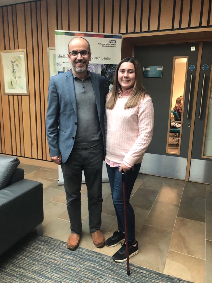
Sheffield Institute of Translational Neuroscience (SITraN)
The Maddi Foundation made a grant to Professor Mimoun Azzouz to investigate gene therapy for SPG15. The research was to evaluate the efficacy of a range of viral vectors in delivering therapeutic normal ZFYVE26 gene to the cells of the brain.
Professor Azzouz was previously Head of Neurobiology at Oxford Biomedica and one of the leading experts in the UK in the use of gene therapy for neurodegenerative diseases. He leads an extensive programme at the Sheffield Institute for Translational Neuroscience (SITraN) aiming to replace or silence faulty genes in forms of neurodegenerative diseases which are caused by a known gene mutation.
Gene therapy technology is currently generating great optimism among patients and their families. However, to develop gene-based therapy for SPG15, further extensive studies are needed to refine our strategy before entering clinical application.
Funds are needed to achieve the following aims:
1) Prepare the therapeutic virus for testing in cells and model systems in the lab to generate a proof-of-concept, a critical step before initiating further pre-clinical work;
2) Prepare the therapeutic virus carrier at the quality acceptable for clinical use in humans;
3) Determine the minimal dose of the carrier that generates efficacy in our animal model;
4) Assess potential adverse effects in a regulatory safety study.
5) Secure the licensing needed to initiate human clinical trials.
Cambridge Institute for Medical Research
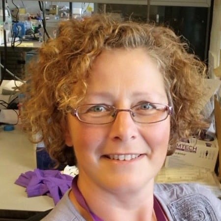
Her focus is understanding the protein machineries that is used by cells to sort cargo proteins for transport to one of a number of destinations. One of these sorting machineries is called AP-5 and involves six proteins working together in a complex – these proteins are the products of the genes SPG15, SPG11, AP5Z1, AP5B1, AP5M1 and AP5S1.
Dr Hirst’s aim is to further understand why faulty versions of SPG11, SPG15 or AP5Z1 have damaging effects on the survival of nerve cells and lead to the rare neurodegenerative disorder ‘hereditary spastic paraplegia’.
Since it is impossible to look inside patient’s nerve cells, cells are taken from patients via a small skin punch that can be preserved and maintained in the laboratory.
Using high powered microscopes the scientists can see that the ‘recycling plant’ compartment of the cell – known as the lysosome – accumulates material that it is seemingly unable to digest. It is believed that this is a build-up of undigested material that is likely to be ‘damaging’ to the health of nerve cells. However, scientists still do not have a clear picture of the precise roles of AP-5 and SPG11 and SPG15.
There have recently been discovered some clues that point to their role in sorting proteins away from the pathway that leads to the lysosome, which Dr Hirst believes would abrogate the damaging accumulation of material in lysosomes. However, there is still a long way to go to understanding the precise function or functions of AP-5, SPG11 and SPG15.


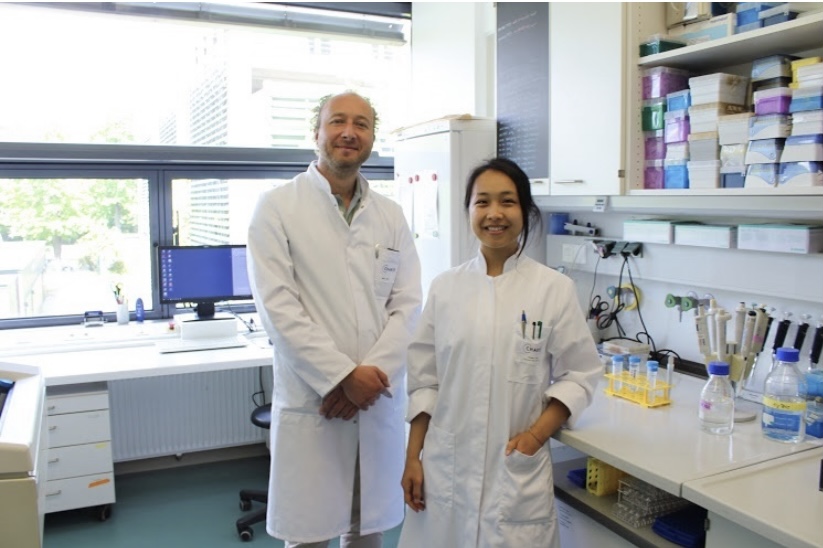
Charite University Berlin Germany
Our research focus is the molecular biology of hereditary neurodegeneration with a focus on “Hereditary Spastic Paraplegias (HSP)”, a group of currently incurable hereditary diseases.
First, our studies aim to identify molecular biomarkers that can be used to develop new laboratory methods for the early diagnosis as well as the therapy monitoring of HSP.
Second, in vivo and ex vivo approaches will define the therapeutic potential of somatic gene repair for HSP.
To this end, the molecular environment of the Zfyve26/Spastizin (HSP type SPG15) protein will be investigated in murine and cell culture models. This will include phenotypic, histological, biochemical and cell biological analyses in combination with (electron) microscopic techniques.


